Spinal Stenosis
Spinal Stenosis
What It Is
In spinal stenosis, exactly what the condition’s name describes occurs: a narrowing of the spinal canal in the cervical, thoracic (more rarely), or lumbar spine, with simultaneous compression of the spinal cord and/or the nerves. Spinal stenosis is a result of spinal degeneration. Patients often present with no symptoms at the early stages of the disease, and what typically triggers symptoms is nerve compression, usually following some form of trauma.
Spinal Stenosis
Symptoms
The symptoms of spinal stenosis, depending on the affected region, may include:
1.
Pain, heaviness, and weakness in the legs that worsen with walking and are relieved when sitting (neurogenic claudication)
2.
Lower back pain that improves with forward bending and rest
3.
Tingling and burning sensation in the lower limbs
4.
Very rarely, patients may experience urinary or bowel dysfunction (incontinence)

Spinal Stenosis
Diagnosis
During physical examination, patients may exhibit weakness and decreased sensation, which are strong indicators that something is wrong. An MRI can show the extent of the damage.
Spinal Stenosis
Treatment
Conservative Management
Many forms of spinal stenosis can be managed with anti-inflammatory medications and physiotherapy. In cases of acute pain, corticosteroids may be prescribed. However, the limitation in mobility and, ultimately, in daily activities usually necessitates surgical treatment.
Surgical Management
When conservative treatment fails, surgical therapy follows, with the goal of decompressing the compressed nerves. How this is achieved depends on several factors, such as the technique and method applied by each surgeon, their surgical skill, and their ability to decompress through small openings rather than large incisions.
More specifically, the following surgical approaches exist:
- Traditional laminectomyis an outdated procedure during which all the posterior elements of the vertebra are removed to create space for the nerves by turning the spinal canal into a cavity. Although this method sufficiently decompresses the neural elements, it leaves the spine unstable (unless spinal fusion is performed simultaneously) and requires a lengthy recovery.
- More modern techniquesfollow the same logic but focus only on the exact point of compression, preserving the elements that do not contribute to nerve compression but support spinal stability. This approach results in a smaller surgical wound compared to traditional laminectomy, typically does not cause instability—but still requires a relatively long recovery (about 6 months).
- Microdecompression via tubular retractoris a surgical approach that likely causes the smallest trauma. With this technique, decompression is achieved through a surgical opening—not an incision—and the working tube passes between the muscles without cutting them. Decompression is performed using a microscope and specialized micro-instruments, and it is possible to decompress the opposite side of the canal from a single approach—even when stenosis is bilateral.
1.
Which surgical method is best and safest for me?
Each case is different, and no single procedure is considered the best for all patients. However, the vast majority of patients benefit significantly from the rapid recovery and short hospital stay—often without overnight admission—allowed by the microdecompression method, which provides equal or even better results compared to more traditional surgical techniques.
2.
How many years of experience does Dr. Starantzis have with this technique?
Dr. Starantzis has extensive experience in the microdecompression technique, having operated on a four-digit number of patients as a lead spine surgeon over the past decade in Greece and the UK.
3.
What kind of anesthesia will I receive?
Spinal stenosis surgeries are usually performed under general anesthesia. However, in selected patients and for operations involving up to two levels, surgery may also be performed WITHOUT GENERAL ANESTHESIA using a specialized protocol of spinal and epidural anesthesia. Dr. Starantzis is one of the pioneering spine surgeons who regularly and successfully applies this protocol in Greece, and the only one performing even spinal fusions without general anesthesia.
4.
How many days will I stay in the hospital?
The microdecompression procedure requires only one day of hospitalization (no overnight stay), in contrast to the classical method, which may involve a hospital stay of up to four days.
5.
When can I return to work?
This depends on the nature of your work. Return to manual labor is allowed after six weeks. If the work is office-based, return is possible after two weeks.
6.
When can I lift weights or exercise again?
As with manual labor, return to sports activities and weight lifting is allowed after six weeks.
7.
What is the success rate of this surgical method?
When applied with the correct indications in the right patients, the success rate exceeds 90–95%.
8.
What are the postoperative complications?
Surgical treatment of spinal stenosis is generally a very safe procedure—among the safest spine surgeries. Nonetheless, there is a minimal statistical risk of complications.
The most serious complication is partial neurological injury, which occurs in fewer than 1% of cases.
Another potential complication, as with all surgeries, is the risk of infection, which is prevented through prophylactic antibiotics.
Rarely, cerebrospinal fluid leakage can occur and may delay wound healing, but statistically, it does not affect the surgical outcome or cause long-term problems.
9.
Could I have a recurrence, and what happens in that case?
There is a possibility of symptom recurrence due to a new manifestation of the disease at a different spinal level. However, if the initial decompression was done correctly, recurrence at the same level is extremely rare.
Endoscopic Decompression
An advanced evolution of tubular microdecompression is endoscopic decompression. In this method, the microscope is replaced by a camera, and the working tube is reduced from 18 mm (typical for tubular microdecompression) to just 10 mm. The surgical trauma is even smaller; however, there is no significant difference in hospitalization or recovery time. When a surgeon is new to this technique or lacks sufficient experience, there is a risk of inadequate decompression and the need for reoperation.
Lumbar Spinal Fusion
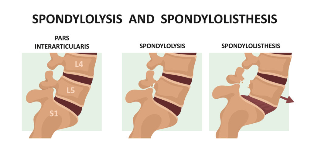
Lumbar fusion is a surgical procedure used to treat instability in the vertebrae (e.g., spondylolysis, spinal stenosis). It is performed by a specialized spine surgeon who inserts rods and screws made from special metal such as titanium.
The ultimate goal is to stabilize the spine and minimize any existing neurological deficit.
Minimally Invasive Transforaminal Interbody Fusion (TLIF)
This is a minimally invasive technique where the spine is operated on percutaneously, without the need for an open approach. The result is less damage to bones and muscles in the area, shorter hospital stay, and rapid recovery.
Minimally Invasive Lateral Fusion
The primary aim of this procedure is to correct spinal curvature and restore alignment using special implants, secured with plates and screws. Bone grafting is also applied.
The operation is performed with a minimally invasive lateral approach (MIS XLIF) at the levels affected by scoliosis and spinal stenosis.
Percutaneous Microdecompression and Minimally Invasive (MIS) Fusion
A combined procedure that can offer significant benefits in spinal stenosis, provided the patient is an appropriate candidate.
Corpectomy
When vertebral body damage is such that it cannot be corrected otherwise and the spinal cord is compressed, the surgeon must intervene decisively by performing a corpectomy. It may be the procedure of choice for fractures, tumors, or cervical myelopathy. It is performed under general anesthesia and is considered a complex and demanding surgery, but one that can prevent disability.
Laminoplasty
Laminoplasty is a technique that effectively treats extensive cervical spinal stenosis and cervical myelopathy, particularly in younger patients.
With this method, a “door-like” mechanism is created at the back of the spinal canal by elevating at least three laminae—similar to opening a door. This expands the spinal canal and allows the spinal cord to revascularize.
It is a clever technique that preserves neck mobility (unlike spinal fusion), relieves spinal cord compression, and allows early patient mobilization.